
The cornea (= cornea) is the shiny, transparent membrane that makes up the anterior part of the eye. Together with the tear film, it serves to protect the eye. In order to understand the formation of the corneal ulcer in a dog, an understanding of the structure of the cornea is necessary.
The cornea consists of several layers. The outermost layer is a so-called multilayer squamous epithelium, which consists of several cell layers. The lowest layer is called the basal membrane. Below this is the corneal stroma consisting of collagen fibers. This is followed by the so-called Endothelium with Descemet’s membrane. The individual layers cannot be differentiated with the naked eye, but can be stained in the event of corneal damage.
Depending on which corneal layer is affected, this is called either superficial corneal erosions/abrasion (= superficial corneal defect that only affects the cell layers of the epithelium) or deeper ulcers (corneal ulcers, plural: ulcers). Once the epithelium is damaged and the Stroma is affected, it is a corneal ulcer in a dog. In the worst case, the ulcer goes down to Descemet’s membrane (= Descemetocele). This condition is extremely critical, as corneal perforation is possible as the course progresses, which can lead to an “lead” of the eye up to an absolute loss of the eye.
How does a corneal ulcer occur?
Most corneal ulcers are traumatic. They are often the result of injury caused by e.g. increased rubbing, cat claws, pointed objects (shorn) or reactions to chemicals, e.g. irritating shampoo. In addition, systemic diseases such as diabetes mellitus, Cushing’s disease or hypothyroidism (underactive thyroidism) can promote the development of corneal ulcers. In some breeds, due to congenital anatomical characteristics (races with prominently protruding eyes, e.g. Pug and French Bulldog) more often come to corneal ulcers. Especially animals with keratoconjunctivitis sicca (KCS, “dry eyes”) often have chronic, poorly healing corneal ulcers. In the boxer, a hereditary corneal weakness is also known, which results in epithelial defects. Medium-aged animals from approx. 6-7 years. In general, however, all breeds of all ages can develop a corneal ulcer.
What symptoms can be found in a corneal ulcer?
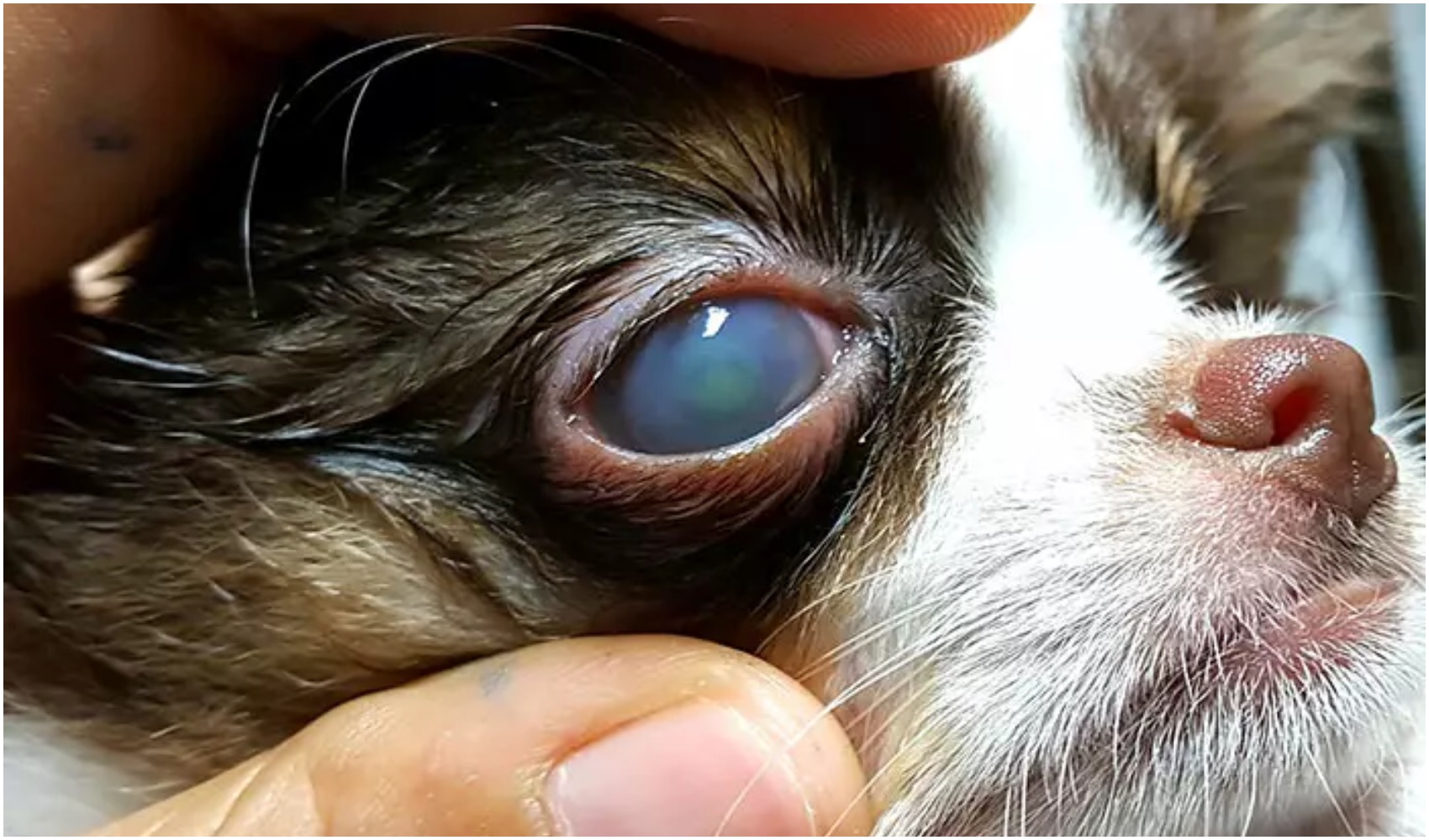
The eyelid bandage skin is reddened, sometimes very strong. A corneal ulcer is accompanied by pain. This means that the dog will try to rub its eye. Very typical is the pain-related pinching of the eyelids (= blepharospasm). There is increased tear flow of the eye up to slimy-purulent eye discharge. Depending on the damage, either a slight turbidity of the cornea can often also be observed with the naked eye.
How is a corneal ulcer in a dog diagnosed?
In addition to a detailed examination of the eye by means of a slit lamp microscope, a staining of the cornea by means of fluorescein is also necessary. The fluorescein sample can be used to stain non-epepilated areas of the cornea and accordingly make a statement about the extent of the damage. In chronic, non-healing defects, swab samples are often obtained for a bacteriological examination.
How is a corneal ulcer treated?
The treatment depends on the severity of the corneal defect. In superficial abrations, vitamin A-containing and/or nourishing eye medications are sufficient to promote epithelization and an analgesic component (usually eye drops containing atropine). As a supportive effect, antibiotic eye drops are usually prescribed to protect against secondary bacterial infections. Especially in the initial phase, a frequent drop is necessary every 4-6 hours.
In the case of deep corneal ulcers, protection of the eye surface is necessary. Since dogs rarely tolerate eye patches, the cornea must be protected by surgical intervention. Here, either the eyelids are temporarily closed (= Tarsorrhaphie) or the nodinal skin is placed over the defect by means of a suture (=nick skin apron). In descemetocele or perforated (=broken) corneal ulcers, a so-called Conjunctival plastic (“flap”) or corneoconjunctival transposition (CCT) is necessary. Here, a portion of the conjunctiva or healthy cornea is sewn onto the affected area in order to achieve the regeneration of the cornea. After healing, the former ulcer will be scarred.
In chronic, non-healing superficial ulcers, on the other hand, it is necessary to remove the dead, poorly healing superficial cornea layers. Your eye specialist will discuss the best therapy option with you.
In general, the earlier the diagnosis is made and therapy initiated, the better the prognosis!
Are there any side effects of the eye drops?
Occasionally, reactions to eye drops/ointments occur. If your dog appears more painful after applying the medication, please contact your veterinarian.
Atropine is often used for pain relief. It enlarges the pupil and can possibly cause photo shyness. The condition is reversible (= reversible) and disappears independently a few days after the end of therapy.
Occasionally, increased salivation and/or smacking/ rubbing of the muzzle is observed after the use of the eye medications. This is due to the fact that a small proportion of eye medications also enter the throat via the tear nose canal. Most eye medications taste bitter, which leads to the described reactions. This condition is short-lived and without hesitation.
What is the course and prognosis for a corneal ulcer in a dog?
For superficial corneal defects, the prognosis for the affected eye is very good. An improvement can be seen just a few days after the start of therapy with appropriate eye medications. In order to prevent a chronic course, a final check by the veterinarian should always be carried out before discontinuation of the eye medication.
As part of the healing process, depending on the depth of the defect, there will be a vascular inciation (= neovascularization) of the cornea, which becomes visible in fine red capillaries in the cornea. These processes speak for a healing ulcer and are desirable.
Chronic defects are often harder to control depending on the cause and have a risk of recurrence (= recurrence of symptoms). Surgical therapy for deep defects also has a good prognosis with the right drip plan that follows an operation.
In surgical care by means of conjunctival apron, turbidity as a result of scarring in the area of the former ulcer is visible after healing. This is the result of the healing process and usually does not affect vision.